Diplomacy fails? Military options off the table? Let’s assassinate the scientists

While I for one do not want Iran to have nuclear weapons I think this is disturbing: Iranian nuclear scientist killed in Tehran explosion – latimes.com
So – basically – the decision by some agency or agencies (US, Israeli, others) seems to be – let’s go after the scientists. Why do they do this? Well, because nobody seems to have the skills or desire to develop an international consensus or diplomacy for an alternative response. Not that dealing with Iran would be easy. And not that military options would be good. But sad in many ways that the option of choice is to kill the scientists behind the work. If all other options had been tried – this might be reasonable – but it does not seem to me that all other options have been tried yet …

Wrap up of tweets from Joe Derisi talk

Just a quick post here – for those not following on Twitter – Joe Derisi gave a talk at UC Davis 1/9 and I posted some tweets about it.  Here they are:

phylogenomics
Joe DeRisi getting ready for his talk at the #ucdavis Genome Center this am http://t.co/OwZbM2nn
1/9/12 9:57 AM
phylogenomics
Joe Derisi’s first slide : “Bees, viruses, and plastids: a seminar in two parts” – I think he needs to work on his math http://t.co/0nPjChlc
1/9/12 10:05 AM

phylogenomics
DeRisi works with mobile honeybee colony trucks that travel around the country to service pollination needs
1/9/12 10:09 AM

phylogenomics
DeRisi – one of the problems with figuring out what is causing CCD is we do not know much about “normal” healthy colonies
1/9/12 10:09 AM

phylogenomics
Bee trucks start in Mississippi in winter, spring in Dakotas, then To california for almonds
1/9/12 10:10 AM
phylogenomics
DeRisi got into bee studies because of colony collapse disorder CCD
1/9/12 10:13 AM

phylogenomics
DeRisi – grinds up bees and then assays them with “bee pathogen chip” and sequencing
1/9/12 10:13 AM

phylogenomics
Derisi discussing his new Plos one w/ SFSU paper showing phorid flies associated with bees
1/9/12 10:16 AM

phylogenomics
Derisi says if you want your paper to get 1000s of hits you should mention Zombies somewhere as they did w/ “Zombie bees”
1/9/12 10:18 AM

phylogenomics
Derisi: zombie bees are cool but the phorids are probably not associated with CCD
1/9/12 10:19 AM

phylogenomics
DeRisi found six major pathogens associated w/ bees: Nosema, phorids, spiroplasma, notovirus, and others
1/9/12 10:22 AM

phylogenomics
DeRisi – sequencing ground up bees – most reads are host or pathogens they know about – says key to characterizing new viruses is assembly
1/9/12 10:23 AM

phylogenomics
DeRisi mentioning their PRICE assembler – an inductive assembly method
1/9/12 10:25 AM
notSoJunkDNA
@phylogenomics URL for PRICE is http://t.co/bRGiY7Rz
1/9/12 10:30 AM

phylogenomics
Why I love Joe Derisi “there will be a manuscript on the assembler someday… But the software is available for free now”
1/9/12 10:27 AM
phylogenomics
DeRisi pooled together Bee samples prepped with various methods so that he could get diverse viruses covered
1/9/12 10:29 AM
phylogenomics
DeRisi – lake sinai virus 2 is present in up to 10^11 copies per bee
1/9/12 10:35 AM

phylogenomics
DeRisi – also present in bees – Crithidia – a trypanosome – related to known pathogens of other insects
1/9/12 10:36 AM

phylogenomics
DeRisi – Crithidia also increases in abundance in winter
1/9/12 10:36 AM
phylogenomics
DeRisi – sequenced and assembled genome of Crithidia – genome is interesting
1/9/12 10:37 AM
phylogenomics
DeRisi – part two – the essential function of the Plasmodium apicoplast
1/9/12 10:38 AM

phylogenomics
DeRisi – history of Studies of plasmodium apicoplast – organelle bound by four membranes – likely b/c result of secondary symbiosis
1/9/12 10:39 AM

phylogenomics
Derisi discussing sequencing of apicoplast genome in 1990s by Gardner, McFadden, etc
1/9/12 10:40 AM

phylogenomics
DeRisi – experiments suggest IPP is the sole essential product of apicoplast biology
1/9/12 10:47 AM

Draft post cleanup #13: Twisted tree of life award: MSNBC, Aliens and Photosynthesis

Yet another post in my “draft blog post cleanup” series.  Here is #13 from July 2010.

I wanted to give this article a “Twisted Tree of Life Award“:

How to find aliens: Follow the photosynthesis – Technology & science – Space – Space.com – msnbc.com

It is pretty painful to me.  Basically the people they are quoting argue that since “complex” life on Earth requires oxygen and since oxygen only comes from photosynthesis, therefore we should look for planets where photosynthesis is possible as the place where life is most likely to be interesting.  Uggh.  So many things in the article I did not like … but just not enough time I guess to bitch about it then.  I will leave it to readers to decide for themselves I guess …

To be discussed in journal club here today: eukaryotic metatranscriptomics

Going to be discussing this in a journal club today

Will post some comments / notes later / as I have them but any comments from others welcome too …
PLoS ONE: Metatranscriptomics Reveals the Diversity of Genes Expressed by Eukaryotes in Forest Soils

Note – am copying the whole text here to mark it up a bit since it it awkward to try and mark it up at the PLoS One site

Metatranscriptomics Reveals the Diversity of Genes Expressed by Eukaryotes in Forest Soils

Coralie Damon1Frédéric Lehembre1,Christine Oger-Desfeux2Patricia Luis1Jacques Ranger3Laurence Fraissinet-Tachet1Roland Marmeisse1*
1 Ecologie Microbienne, UMR CNRS 5557, USC INRA 1193, Université de Lyon, Université Lyon 1, Villeurbanne, France, 2 Pôle Rhône-Alpes de Bioinformatique, Université de Lyon, Université Lyon 1, Villeurbanne, France, 3 Biogéochimie des Ecosystèmes Forestiers, INRA centre de Nancy, Champenoux, France

Abstract Top

Eukaryotic organisms play essential roles in the biology and fertility of soils. For example the micro and mesofauna contribute to the fragmentation and homogenization of plant organic matter, while its hydrolysis is primarily performed by the fungi. To get a global picture of the activities carried out by soil eukaryotes we sequenced 2×10,000 cDNAs synthesized from polyadenylated mRNA directly extracted from soils sampled in beech (Fagus sylvatica) and spruce (Picea abies) forests. Taxonomic affiliation of both cDNAs and 18S rRNA sequences showed a dominance of sequences from fungi (up to 60%) and metazoans while protists represented less than 12% of the 18S rRNA sequences. Sixty percent of cDNA sequences from beech forest soil and 52% from spruce forest soil had no homologs in the GenBank/EMBL/DDJB protein database. A Gene Ontology term was attributed to 39% and 31.5% of the spruce and beech soil sequences respectively. Altogether 2076 sequences were putative homologs to different enzyme classes participating to 129 KEGG pathways among which several were implicated in the utilisation of soil nutrients such as nitrogen (ammonium, amino acids, oligopeptides), sugars, phosphates and sulfate. Specific annotation of plant cell wall degrading enzymes identified enzymes active on major polymers (cellulose, hemicelluloses, pectin, lignin) and glycoside hydrolases represented 0.5% (beech soil)–0.8% (spruce soil) of the cDNAs. Other sequences coding enzymes active on organic matter (extracellular proteases, lipases, a phytase, P450 monooxygenases) were identified, thus underlining the biotechnological potential of eukaryotic metatranscriptomes. The phylogenetic affiliation of 12 full-length carbohydrate active enzymes showed that most of them were distantly related to sequences from known fungi. For example, a putative GH45 endocellulase was closely associated to molluscan sequences, while a GH7 cellobiohydrolase was closest to crustacean sequences, thus suggesting a potentially significant contribution of non-fungal eukaryotes in the actual hydrolysis of soil organic matter.

Introduction Top

In terrestrial ecosystems, plant litter degradation is a key ecological feature which controls not only the equilibrium between soil carbon storage and CO2 release in the atmosphere, but also the release of essential soil nutrients trapped in dead plant biomass such as organic forms of phosphorus and nitrogen.
In ecological studies, litter degradation is often estimated by measuring parameters such as soil respiration [1], litter mass loss [2] or the activities of specific microbial enzymes in soil extracts [3]. In microbiology, the degradation of plant-derived compounds such as lignocellulose has been studied using a few microbial model species and has recently led to the sequencing of the genomes of different saprotrophic fungal species which use different strategies to degrade plant material [4][7], thus revealing the full enzymatic machinery implicated in this process.
Under natural conditions, litter degradation is generally carried out by consortia of species that either act simultaneously or replace one another on a common piece of plant debris in a sometimes predictable manner and not by a single microbial species [8]. It can therefore be anticipated that the molecular machinery deployed to completely mineralize litter in the field is far more complex and diverse than the machinery observed in a single microbial genome. In addition, it is likely that the diversity of this machinery is partly controlled by litter chemistry and complexity and therefore by plant community composition. Indeed, by affecting litter quality and soil physicochemical properties, plant cover could influence microbial community composition and/or diversity [9][10], and select different microbial taxa that may possess different degradation machineries.
By allowing access to the genome contents of the different microorganisms present in a common environment (metagenomics) or to the set of genes they express (metatranscriptomics), environmental genomics offers a novel opportunity to decipher at the molecular level, complex ecological processes such as plant organic matter degradation, thus bridging the gap between global field measurements and targeted genomic approaches. These approaches have been carried out on a variety of animal digestive systems [11][17] or model composts [18]and DNA and/or RNA sequencing of their associated microbial communities has lead to the characterization of numerous members of carbohydrate-active enzyme families whose frequencies differ depending on the origin of the sequenced metagenomic DNA/RNA [11],[15].
Since the primary degradation of lignocellulosic materials in soil is believed to be carried out essentially by fungi, we present a first metatranscriptomic analysis of forest soils focussing specifically on eukaryotic organisms whose polyadenylated mRNA can be separated from prokaryotic and non coding ones, thanks to their poly-A tails [13],[19][20]. Poly-A mRNA were directly extracted from beech (Fagus sylvatica, a broadleaf deciduous angiosperm species) and spruce (Picea abies, an evergreen gymnosperm species) forest soils, converted into cDNA then cloned and 10,000 of them per library sequenced using the Sanger technology. The sequence datasets were specifically analysed with respect to the diversity of enzymes implicated in the breakdown of lignocellulosic material and downstream monosaccharide membrane transporters, as well as enzymes and transporters involved in the mobilization of the organic forms of other elements (phosphorus, nitrogen). In addition, since many microbial enzymes produced for soil nutrient acquisition, and/or toxic soil compounds inactivation (e.g. P450 monooxygenases) are also of biotechnological interest, we performed a global evaluation of the sequence dataset with respect to its biotechnological potential.

Results Top

Sequence datasets (Table 1)

thumbnail

Table 1. Characteristics of the sequence datasets.

doi:10.1371/journal.pone.0028967.t001

cDNA libraries of ~9 105 and 5 105 plasmid clones were obtained using poly-A mRNA (NOTE – THEIR USE OF polyA FOCUSED PREP WILL DEFINITELY BIAS THIS IN FAVOR OF EUKS THOUGH WILL ALSO MISS SOME THIGNS)  extracted from beech and spruce soils, respectively. A significant proportion of cDNA inserts were between 400 and 700 bp-long, as estimated by agarose gel electrophoresis (64% and 48% of cDNA for beech and spruce respectively). Following sequencing and cleaning of the sequences (removing bad quality sequences, poly-A tails, vector and adaptor sequences as well as contaminating rRNA sequences (NOTE – NOT SURE WHY THEY REMOVED rRNA), and discarding sequences shorter than 100 nt), 7905 (beech) and 8606 (spruce) cDNAs were retained for further analyses. Contamination levels by eukaryotic and bacterial rRNA sequences represented at most 8.8% of the sequences (beech). These rRNA sequences could have been cloned because they processed A-rich sequence stretches, and therefore may not reflect the taxonomic diversity of the soil microbial communities. Finally the average read length was 430 bp for beech and 482 bp for spruce cDNAs.
For spruce and beech cDNA sequence datasets, 9% of the sequences grouped into clusters which, for more than 70% of them, contained only two sequences (Figure S1 B–C). As a result, the rarefaction curves plotting the numbers of clusters versus the numbers of sequences had the shapes of straight lines (Figure S1 A) suggesting that the datasets represented a small proportion of the total metatranscriptome sequence diversities. Only two of the 12 clusters grouping at least 8 sequences corresponded to known genes. One, from the beech library, encoded a fungal hydrophobin, a class of proteins which coat the external surface of hyphae and spores, and the other, from the spruce library, a putative chitinase.

Taxonomic diversity of the soil eukaryotic communities

For each forest soil, the taxonomic composition of the eukaryotic community contributing to the soil RNA pool was independently estimated using two sequence datasets: (i) 18S rDNA fragments PCR amplified from reverse-transcribed soil RNA (Table S1), and (WHY NOT ALSO LOOK AT THE rRNAs THEY RECOVERED IN THE cDNA LIBS?) (ii) the sequenced cDNAs. Sequences were assigned to one of eight major eukaryotic phyla, i.e. the Fungi, Metazoas, Plantae, Amoebozoas, Alveolata, Heterokonta, Rhizarias and Excavata (as defined in [21]). The last 5 phyla are collectively referred as the “protists” in the manuscript. NOT SURE THIS IS A GOOD USE OF THE TERM.
Whatever the sequence dataset and the studied soil, the Opisthokont group (i.e. essentially Fungi and Metazoas) dominated, representing 71% (beech) and 77% (spruce) of the unambiguously annotated PCR amplified 18S rDNA and 60–61% of the annotated cDNAs (Figure 1 andTable S2). Furthermore, among the Opisthokonts, fungal sequences were the most abundant, representing between 75 and 87% of the cDNAs attributed to this group.
thumbnail

Figure 1. Taxonomic affiliation of the different environmental sequences.
PCR amplified 18S rRNA (column 1) and cDNAs (columns 2 and 3) from spruce and beech soils were attributed to one of eight eukaryotic phyla or to the Bacteria. The “multiple affiliation” category contains sequences which could not be unambiguously placed in one of the other categories. (A), Results for all sequences which had a homolog in the GenBank/EMBL/DDJB database; (B), same analysis after removing cDNA sequences from bacteria and cDNA with uncertain “multiple affiliation” for a direct comparison between 18S rRNA and cDNA datasets. For the annotation of cDNA sequences using MEGAN, two different parameters were used; the “stringent analysis” (column 2) corresponded to the “Min Support” parameter set to default value of 5 which was set to 1 in the “less stringent” analysis (column 3).

doi:10.1371/journal.pone.0028967.g001

Sequences from each “protist” group were identified at least once in each of the different beech or spruce sequence datasets and protist sequences contributed to the different datasets between 3% (cDNAs) and 12% (amplified 18S rDNA) of the unambiguously annotated sequences. It should however be stressed that the use of a single primer pair to amplify eukaryotic rDNA genes is known to capture only a fraction of the true diversity [22] and that amplification of 18S sequences from several groups of unicellular eukaryotes requires group-specific primers [23].
Using the default parameters and the BLASTX results against the NCBI-nr protein database as input file, the MEGAN software attributed a taxonomic annotation to 46% (spruce) and 37% (beech) of the cDNAs. Among these annotated cDNAs, only 2.5% were attributed to “protists” and 22–29% could not be unambiguously attributed to a single taxonomic group (multiple affiliation category, Figure 1). We asked if under-representation of specific taxonomic groups, such as the protists, and a high prevalence of sequences with multiple potential affiliations may have resulted from the settings of the MEGAN software. Indeed, using default parameters, MEGAN could take into consideration a too high number of BLAST hits (a maximum of five) to infer a taxonomic affiliation thus underestimating the abundance of taxonomic groups poorly represented in sequence databases. This hypothesis was tested by lowering the “Min Support” parameter from 5 to 1 (relaxed setting). Although this modification increased the number of sequences attributed to protists by 50% (which still represented at most 3.5% of the cDNAs); this did not change the overall taxonomic distribution of the cDNAs (Figure 1).

Global functional annotation of the cDNAs

Using an e-value threshold of 10−6, 48% (spruce) and 39.5% (beech) cDNA sequences returned a positive hit in BLASTX searches against the GenBank nr protein database. Among the sequences with no homolog in this preliminary analysis, an additional 227 (spruce) and 238 (beech) contained a registered protein motif identified using InterProScan.
Using Blast2GO, a Gene Ontology (GO) term (associated to either a biological process, a molecular function or a cellular component GO category) could be assigned to 31.5% (beech) and 39% (spruce) of the cDNAs. The five most abundant “biological process” and “molecular function” GO categories were the same for the beech and spruce datasets and altogether accounted for respectively 44% and 43% of the sequences annotated with either a “biological process” and “molecular function” GO term (Figure S2). The two most abundant categories were related to protein synthesis (ribosome biogenesis and translation) and contained, for example, ribosomal proteins encoding sequences.
Finally, 12–13% of the sequences in each datasets encoded proteins with enzymatic activities to which an Enzyme Commission (E.C.) number could be associated (Table 1). Two hundred different E.C. numbers were placed into 117 (spruce) and 112 (beech) different KEGG pathways. Several of these pathways such as glycolysis, tricarboxylic acids (TCA) cycle, nitrogen and sulfur metabolisms, amino acid biosynthesis or degradation (Table 2) are essential to appreciate basic soil processes such as global microbial activities and soil nutrient assimilation. For several of these pathways we identified key enzymes that represent the obligate entry points of major soil nutrients in fungal metabolism (Table S3). This is for example the case of the glutamine synthase (E.C. 6.3.1.2)/NADPH-glutamate dehydrogenase (E.C. 1.4.1.4)/glutamate synthase (E.C. 1.4.1.13) for ammonium; the adenylyl-sulfate kinase (E.C. 2.7.1.25) and the sulfate adenylyltransferase (E.C. 2.7.7.4) for sulfate and aldehyde reductase (E.C. 1.1.1.21) for pentoses (xylose, arabinose) which are end products of hemicelluloses hydrolysis (Table S3).
thumbnail

Table 2. A list of KEGG pathways relevant to soil nutrient (C, N, S) utilisation and microbial metabolism for which different cDNA sequences from the spruce and beech soil could be affiliated.

doi:10.1371/journal.pone.0028967.t002

Targeted annotation of the cDNAs

Our annotation effort concentrated on gene categories encoding proteins with biochemical activities directly linked to the breakdown of plant compounds and other complex nutrients (lignocellulose, chitin, proteins but also secondary metabolites) and polypeptides involved in the mobilisation of soil nutrients such as transmembrane transporters. This targeted annotation was essentially performed through sequence homology searches against specialized protein databases, which compile members of each of these different protein categories, or by using reference sequences when such specialized databases did not exist (in the case of phytases and dioxygenases). Results were further filtered manually and/or by using the MetaBioME database [24] to remove enzymes with no obvious links to the studied processes (such as for example intracellular proteases component of the proteasome).
This annotation strategy identified a large variety of enzymes involved in the above-mentioned processes including many different enzymes classes active on most plant cell wall polymers (cellulose, hemicellulose, pectin and lignin) but also on starch and chitin which, as a component of arthropods exoskeleton and fungal cell wall, represents an abundant nitrogen source in forest soils (Table 3). Altogether, Glycoside Hydrolases (GH CAZymes) represented between 0.5% (beech) and 0.8% (spruce) of all cDNAs. In addition we also identified a set of enzymes responsible for the breakdown of non-cell wall organic molecules such as different classes of proteases, a phytase, a cutinase and a putative carotenoid ester lipase (Table S4). These latter enzymes were all highly similar to known excreted fungal enzymes.
thumbnail

Table 3. An illustration of the diversity of potential plant cell wall and other polysaccharides (starch, chitin) degrading enzymes identified among the beech and spruce soil ESTs.

doi:10.1371/journal.pone.0028967.t003

Given that organic matter input in soil also results in the input of toxic secondary metabolites, we searched for enzymes classes potentially involved in their detoxification. While we only identified a single putative fungal dioxygenase (cathechol dioxygenase) among sequences from the spruce soil, we identified a total of 26 P450 monooxygenases belonging to 12 different families (as defined in the CYPED [25]database, Table S4). Among the CYPED homologous sequences, several did indeed contribute to the breakdown of toxic compounds such as benzoate or the flavonoid phytoalexin pisatin while others were part of biosynthetic pathways such as an O-methylsterigmatocystin oxidoreductase involved in aflatoxin production.
Besides their involvement in organic matter processing, most of these different enzymes are targets for the bioindustry. We therefore performed a global annotation of the environmental EST datasets with respect to their content in genes of biotechnological interest by blasting all sequences against the MetaBioME database which compiles sequences of “Commercially Useful Enzymes” (CUE). For both spruce and beech soils, more than 3% of the sequences returned a positive hit in this analysis and the two most represented CUE classes were the oxidoreductases and the hydrolases (Figure S3). About half of these sequences were from fungi (Figure S3).
Both spruce and beech soil metatranscriptomes included significant proportions (6.2 and 4.8% respectively; E-value threshold of 10−6) of putative membrane transporters as identified by blasting all cDNA sequences against the Transporter Classification Database (TCDB;Table 4[26]. The most abundant category of fungal transporters involved in the assimilation of soil nutrients was the sugar porter one (T.C. 2.A.1.1) implicated in both hexoses (eg glucose, galactose, mannose) and pentoses (eg arabinose, xylose) assimilation, followed by the different families participating to amino acid and phosphate uptake. Concerning inorganic nutrients, besides phosphate transporters, we also identified ammonium transporters (T.C.1.A.11.) but neither nitrate (T.C.2.A.1.8) nor sulfate (T.C.2.A.53.1) ones.
thumbnail

Table 4. Diversity of fungal plasma membrane transporters, potentially involved in soil nutrient uptake, identified among the beech and spruce soil cDNAs.

doi:10.1371/journal.pone.0028967.t004

For most of the explored gene categories we consistently identified a higher number of homologous sequences in the spruce dataset compared to the beech one (Tables 3S3 and 4). The most striking difference concerned the sugar porter family with 31 sequences in the spruce forest metatranscriptome (0.34% of the sequences) against only one in the beech forest data set (0.01%).

Identification of full-length CAZymes

Twelve presumably full-length Carbohydrate Active Enzymes (CAZyme) encoding cDNAs, belonging to families CE1 (accession no. FR865947), GH5 (FR865941), GH7 (FR865943-4), GH11 (FR865942), GH45 (FR865945), GH61 (FR865936-40) and PL1 (FR865946), were identified and sequenced entirely. The longest cDNA clone was 1671 bp-long and encoded a putative GH7 cellobiohydrolase. Except for the CE1 family member, which is thought to be an intracellular enzyme with S-formyl glutathione hydrolase activity, all other 11 predicted protein sequences possessed a N-terminal signal peptide in agreement with the potential polysaccharide degrading activities of the corresponding enzymes.
The putative taxonomic/phylogenetic origin of these 12 cDNAs was assessed using Bayesian (MrBayes) and/or maximum likelihood (PhyML) phylogenetic analyses of the deduced amino acid sequences. Protein sequences used in the phylogenetic analyses included not only the CAZyme best BLASTX hits from GenBank nr database but also homologous sequences identified among GenBank non fungal ESTs in addition to the gene models predicted from 57 recently released fungal and non fungal (animals, choanoflagellate) genome sequences (Table S5).
Each of the 12 full-length environmental sequences were homologous to fungal sequences and to sequences from other taxonomic groups either closely related to the Fungi, such as the Metazoa (GH7 and GH45) or the choanoflagellates (CE1), or to more distantly related groups such as the Plantae (GH5, PL1) or the Bacteria (GH5, GH11 and PL1). With the exception of the Carbohydrate Esterase family 1 (CE1) genes, all other studied CAZyme genes were not present in all genomes analysed and, when present, frequently occurred as gene families from which between one and three members per species were used for the phylogenetic analyses.
In many cases, sequences from one specific taxonomic group (eg Fungi or Bacteria) did not group together to form a single homogeneous and statistically well-supported clade (Figure 2Figure S4). This could be due to an insufficient number of phylogenetically-informative characters in the sequences or to a complex evolutionary history of these gene families characterised by frequent gene duplications, gene losses and potential horizontal gene transfers between distantly related taxonomic groups [27]). In fine, only the phylogeny of the CE1 family fitted to the overall phylogeny of the Fungi [28] and the CE1 environmental sequence most likely originated from an Ascomycota Pezizomycotina fungal species (Figure S4D). Several other environmental sequences, including putative pectin lyase (PL1, Figure S4A), xylanase (GH11, Figure S4B), cellobiohydrolase (GH7, Figure 2B), endocellulase (GH45, Figure 2A) and members of the copper-dependent oxidase GH61 family [29] (Figure S4E), did not group with other referenced sequences to form statistically well supported phyla. This suggests that these sequences originated from fungal or non-fungal taxonomic groups distantly related to those from which homologous sequences have already been characterised. Percentages of identical amino acid positions between the environmental sequences and their closest relatives in the phylogenetic analyses (Figures 2 andS4) supported this hypothesis for several sequences. These percentages ranged from as low as 35–38% for two GH61 and the PL1 sequence to 67–70% for a GH7 and the CE1 sequences (Table S6).
thumbnail

Figure 2. Putative phylogenetic origins of three environmental Glycoside Hydrolases belonging to families GH7 (A) and GH45 (B).
Bayesian (MrBayes) phylogenetic trees include protein sequences from different taxonomic groups, each identified by a specific colour. Diagrams drawn to scale illustrate the modular structure of each of the different GH45 protein sequences. Red rectangles, potential signal peptides; blue rectangles, GH45 domains used for the phylogenetic analysis; orange rectangles, family one Carbohydrate Binding Modules (CBM1) characteristic of fungal GH45; purple rectangle, CBM2 module only found in the environmental sequence. Boxed species names indicate protein sequences for which an endoglucanase catalytic activity has been experimentally established [30][33]. Posterior branch probabilities above 0.8 are given; branches with less than 0.5 probability support were collapsed.

doi:10.1371/journal.pone.0028967.g002


NOTE – I DID NOT LIKE THE GROUPINGS THEY MADE ON THESE TREES – THEY LABELLED AS “GROUPS” POLYPHYLETIC LINEAGES GIVING A DECEPTIVE IMPRESSION THAT THEY ARE ACTUAL PHYLOGENETIC GROUPS
Two of the environmental CAZymes potentially originated from the soil fauna. This was the case for one of the two analysed GH7 (putative cellobiohydrolase Env-GH7/16YJ11; Figure 2A) family members that grouped with the recently identified crustacean cellobiohydrolase polypeptides. This was also the case for the GH45 family member encoding a putative endocellulase (Figure 2B). This latter environmental sequence grouped with Molluscan GH45 sequences but differed from these sequences by the presence of a family two carbohydrate binding module (CBM2) at its C-terminal end (Figure 2B). Carbohydrate binding modules existed in several fungal GH45 proteins but they all belonged to the unrelated CBM1 family.

Discussion Top

This report on the systematic sequencing of environmental eukaryotic cDNAs establishes the main features of this experimental approach. First, the specific analysis of polyadenylated mRNA allows access to the protein-coding gene pool of eukaryotes and efficiently counter-selects not only rRNA but also “non-eukaryotic” mRNA. This is in contrast to the studies published so far on the analysis of “global, essentially prokaryotic, metatranscriptomes” [34][38] which result in sequence datasets comprising as little as less than 1% [35][36] to often more than 50% of rRNA sequences despite the use of different protocols to eliminate these molecules [39][40]. Furthermore, with less than 3% of cDNAs attributed to bacteria in both beech and spruce soil metatranscriptomes, polyadenylation of bacterial mRNA [41] does not seem to represent a challenge for the study of eukaryotic environmental mRNA. Moreover, the exact taxonomic origin of cDNAs attributed to bacteria should to be carefully evaluated as they may include genuine eukaryotic genes not yet identified in this taxonomic group such as bacterial genes recently acquired by eukaryotes by horizontal gene transfer; as for example recently suggested for nematodes carbohydrate active enzymes (CAZymes; [27]).
Secondly, among the cDNAs, between 52 and 60% have no homologs following BLASTX searches against the GenBank nr database. These figures, similar to the one reported for the Muskoxen rumen eukaryotic metatranscriptome [13], are higher than the percentages of orphan genes revealed in recently published genomic sequences from fungi and invertebrate phyla, that dominate the two studied soil eukaryotic communities. Figures vary widely from as low as roughly 5% for the entomopathogenic Ascomycota fungi Metarhizum sp. [42] to 20% for the symbiotic species Tuber melanosporum [43]. In the case of the insect Pediculus humanus [44] and the crustacean Daphnia pulex [45], two arthropods distantly related to other fully sequenced species from this phylum, the percentages of genes without homologs have been estimated to be of 10 and 36% respectively. Factors susceptible to accentuate the low percentage of cDNA taxonomic affiliation in eukaryotic metatranscriptomes include the presence of poorly studied protist groups in soils and also the large proportion of short sequences located at the 3′ end of the cDNAs which comprise untranslated 3′ UTRs. Future use of pyrosequencing or of other high-throughput sequencing technologies may increase the percentages of short reads, a factor that will be mitigated by a sharp increase in the number of sequences produced. All these figures must however be considered as transitory. Indeed, the current exponential increase in the number of available genome sequences in conjunction with a broader taxonomic sampling of the sequenced taxa, more representative of the soil biota, should have a direct and rapid positive impact on the taxonomic annotation of eukaryotic environmental sequences. More specifically it will allow us to carefully evaluate the contribution of protist taxa to the soil metatranscriptomes. Regarding functional annotation, better assignation will depend upon both (i) functional studies on individual unknown environmental sequences [46] and (ii) functional validation of unknown proteins from model organisms.
Despite the large proportion of housekeeping genes involved in basic cell maintenance mechanisms, both global (KEGG pathways) and targeted annotation identified a large variety of genes of interest in a context of soil ecosystem functioning and more specifically in the turnover of plant biomass and soil nutrient cycling and utilization. These nutrients encompass inorganic (ammonium, phosphates) and organic (aminoacids, peptides, phytate) forms of nitrogen and phosphorus, sulfates and simple sugars resulting essentially from either rhizodeposition or lignocellulose breakdown. We also noticed that genes representative of other key pathways such as the nitrate assimilation one (which comprises specific transporters, nitrate and nitrite reductases) were however missing in the datasets which could indicate that the corresponding nutrient represents a minor N source in the studied acidic forest soils.
Enzymes participating to plant cell wall deconstruction are of special interest not only from an ecological point of view to quantify and understand soil C turnover, but also because they are among the most widely used enzymes in the industry and are instrumental to the development of second generation biofuels. We identified among the beech and/or spruce soil cDNAs representatives of enzymes active on the main plant cell wall polymers: lignins, cellulose, hemicelluloses and pectins. Genes encoding carbohydrate active enzymes clearly outnumbered those encoding enzymes directly active on lignin although members of the three lignin oxidase families were identified in at least one of the two metatranscriptomes (Table 3). Glycoside Hydrolases (GH) encoding genes represented 0.46% (beech) and 0.78% (spruce) of the analyzed cDNAs. These figures are very similar to those reported for different “carbohydrate-adapted” metagenomes [18]. In a perspective of mining soil eukaryotic metatranscriptomes for such enzymes these values signify that the screening effort could be similar to the screening effort developed for metagenomes [14]. Additional sequencing effort as well as the study of replicate soil samples are nevertheless required before concluding on a potential effect of litter quality (beech versus spruce) on the relative abundance of transcripts related to plant cell wall degradation or nutrient assimilation (eg sugar transporters). It is indeed known that these two species differ with respect to the nature and proportions of polymers present in their cell walls [47][48] and that spruce litter mineralizes more quickly compared to the beech one [49].
The use of Sanger sequencing of cloned cDNAs allowed us to easily identify several full-length genes among which several CAZyme ones. Phylogenetic analysis of the coded protein sequences suggests that many of them are distantly related to protein sequences present in databases. This further emphasizes the interest of eukaryotic metatranscriptomes as a source of novel enzymes with potentially interesting catalytic properties. This also indicates that organisms studied thus far at the molecular level in a context of plant cell wall deconstruction may not be representative of the organisms at work in the soil ecosystem. It is particularly interesting to consider that at least two out of the 12 full-length CAZymes (putative cellobiohydrolase and endocellulase) could originate not from fungi but from animals. A number of recent publications have indeed reported the existence of plant cell wall degrading enzyme genes in the genome of different groups of invertebrates which are represented among the soil fauna[27][31][33][45][50][51]. This could indicate that in addition to their well recognized role in the fragmentation and mixing of plant litter, the soil fauna could also play a direct and potentially major role in the actual hydrolysis of plant carbohydrates in soils which adds up to hydrolysis by their gut microbiomes.
In conclusion, this first report of a eukaryote-specific analysis of soil metatranscriptomes highlights the potential of this experimental approach in two research fields. In the field of environmental biotechnology, eukaryotic metatranscriptomes represent diversified multigenome resources for many different gene categories used in the bioindustry. In this context, construction of environmental cDNA libraries whose potentially large inserts can be sequenced (this study) or directly expressed in heterologous hosts [20][46][52][53]potentially represents the best experimental approach. In the field of environmental sciences, the study of eukaryotic metatranscriptomes will help us to reevaluate the contribution of the different soil eukaryotes to basic and essential soil processes such as organic matter degradation. It will also contribute to compare ecosystems which differ with respect to i.e. vegetation type, soil characteristics and climate and evaluate the relative contribution of these different variables on different soil processes. In this respect, high-throughput sequencing technologies should be implemented.

Materials and Methods Top

Study site and soil sampling

Soil samples were taken from an environmental research observatory forest site located in central France (Breuil-Chenue forest, 47°18′10″N, 4°4′44″E, 638 m above sea level). This site, initially covered by a mixed broadleaved tree forest (Fagus sylvaticaQuercus sessiliflora,Betula verrucosaCorylus avelana) was clear-cut and replanted in 1976 by separate, single forest tree species stands, among which spruce (Picea abies) and beech (Fagus sylvatica). On July the 10th 2007, 14 (spruce stand) and 16 (beech stand) soil samples (8 cm in diameter, 15 cm in depth) were collected along a systematic sampling grid. For each stand, the uppermost 3–7 cm thick organic matter-rich horizon of each core was sieved (2 mm mesh size) and mixed together (100 ml per core) to obtain a composite sample which was immediately frozen at −70 °C. Pedoclimatic parameters, soil characteristics and sampling strategy are detailed in Table S7.

RNA extraction, cDNA libraries construction and sequencing

Total RNA (~100 µg) was extracted from ~90 g of each of the two composite forest soil samples as described in [20] and [54]. Polyadenylated mRNA were separated from non-polyadenylated RNAs (rRNAs, mitochondrial and prokaryotic mRNAs, tRNAs) by affinity capture on paramagnetic beads coated with poly-dT (Dynabeads Oligo (dT) kit, Dynal). Unbound non-polyadenylated RNAs were recovered by ethanol precipitation. Reverse transcription was carried out following the SMART cDNA Library Construction Kit instructions (Clontech), using between 90 (beech) and 210 ng (spruce) of purified mRNA. cDNA were further amplified by long distance PCR (LD-PCR) as described in the SMART cDNA Library Construction Kit, by using 20 and 24 PCR cycles for spruce and beech respectively. After size-fractionation to remove cDNA smaller than 400 pb (CHROMA SPIN-400 Columns, Clontech), the cDNAs were digested with Sfi1 and ligated into the Sfi1-digested pDNR-LIB plasmid vector (Clontech).
Each cDNA library was introduced in electro-competent E. coli cells (DH10B, Invitrogen) and 10,000 randomly selected cDNA clones were sequenced from their 5′ end using the M13-20 primer and Sanger chemistry (Genoscope sequencing centre, Ivry, France).

18S rDNA gene libraries construction and sequencing

350 (beech) and 730 (spruce) ng of soil non-polyadenylated RNA were reverse transcribed using 0.2 µg of random hexamers and 200 U of M-MuLV Reverse Transcriptase according to the manufacturer instructions (MBI Fermentas). A ca 560 bp-long fragment located at the 5′-end of the eukaryotic 18S rDNA gene was amplified by PCR using primers Euk1A (CTGGTTGATCCTGCCAG) and Euk516R (ACCAGACTTGCCCTCC) described by [55]. PCR mixtures (25 µl) contained 200 nM of each primer, 200 µM of each dNTP, 1 mM MgCl2, 0.25 mg.ml−1 of bovine serum albumin, 0.625 U of Pfu DNA polymerase, the appropriate buffer (Fermentas) and one tenth of the reverse-transcription mix. Amplification reaction started with an initial denaturation step of 3 min at 96°C, followed by 25 cycles comprising 45 sec at 96°C, 45 sec at 56°C and 2 min at 72°C, and finished with an elongation step of 5 min at 72°C. Amplification products of the expected size from 15 PCR tubes were pooled, isolated from an agarose gel (Nucleospin Extract kit, Macherey-Nagel) and ligated in the plasmid pCR-Blunt II-TOPO (Zero Blunt TOPO PCR Cloning kit, Invitrogen) that was used to transform electro-competent DH10B E. coli cells (Invitrogen).
For each rDNA library, the ca 560 bp rDNA inserts of 96 clones were entirely sequenced (Agowa Company, Berlin, Germany) using universal primer M13-20. Sequences were manually corrected and edited. BLASTn searches were performed against GenBank nr nucleotide database at NCBI (http://www.ncbi.nlm.nih.gov/) using default parameters except for word size which was set to 7. Sequences were analyzed with the rRNA Database Project CHECK_CHIMERA program (http://rdp8.cme.msu.edu/). Potential chimeras were further analysed by blasting separately the two dissimilar segments of the sequences against GenBank. Confirmed chimeras and artefacts were eliminated from the sequence datasets. All remaining sequences were submitted to EMBL and are available under accession numbers FN393180–FN393221 (beech) and FN393323–FN393380 (spruce).

cDNA sequences cleaning and clustering

All cDNA sequences were trimmed using TIGR SEQCLEAN tool (http://www.tigr.org/tdb/tgi/software) with default parameters to eliminate poly-A sequences at the 3′end of cDNA, vector, adaptor and primer sequences and undetermined nucleotides. Sequences shorter than 100 nucleotides were eliminated. Contaminant ribosomal RNA sequences were identified by BLASTN analysis (cut-off threshold of E-value≤10−10, word-size = 11) of all sequences against LSUrdb and SSUrdb_SSURef100 libraries described in [38] (available athttp://services.cbu.uib.no/supplementary​/community-profiling/), as well as against NCBI-nr nucleotide database (looking for “ribosomal RNA” or “rRNA” in the title). The resulting “cleaned” sequence dataset was submitted to EMBL and is available under accession numbers FR706059–714330 (beech) and FR697056–706058 (spruce).
Cleaned sequences were clustered using Cd-hit (http://cd-hit.org[56]) using a 90% identity threshold. The resulting clusters were used to perform a rarefaction analysis using S. Holland’s Analytical Rarefaction version 1.3 software (http://www.uga.edu/strata/software/).

Global cDNA sequences annotation

Cleaned cDNA sequences were queried (BLASTX) against NCBI nr protein database (http://www.ncbi.nlm.nih.gov/), using 2 different strategies, (i) Q-BLAST via BLAST2GO [57] with parameters, E-value≤10−6 and overlapping length >33% to the corresponding best hit and, (ii) NetBlast 2.2.22 (E-value≤10−6). As both analyses gave similar percentages of annotated cDNA sequences, we only considered the Q-BLAST results. In addition to raw BLASTX analyses, we also used BLAST2GO to perform Gene Ontology (GO, [58]), Enzyme code (E.C.) and InterPro (conserved patterns in sequences) annotations.

Targeted annotation of cDNAs

Cleaned sequence datasets were searched for sequences similar to genes coding for organic matter degradation enzymes and for enzymes of biotechnological interest, using BLASTX searches (default parameters) against various specialized databases. Searches were made against Carbohydrate Active Enzymes (CAZy,http://www.cazy.org/[59]); and Fungal Oxidative Lignin Enzymes (FOLy; http://foly.esil.univ-mrs.fr/[60]) databases to find plant cell wall degradation enzymes. cDNA similar to cytochrome P450 potentially involved in detoxication of plant secondary metabolites were searched against CYPED database (downloaded on October 2009,http://www.cyped.uni-stuttgart.de/[61]). Lipases/esterases and proteases were searched against the Lipases Engineering database (LED, downloaded on December 2009; http://www.led.uni-stuttgart.de/[62]) and MEROPS (downloaded on January 2010,http://merops.sanger.ac.uk/index.shtml[63]) respectively. Membrane transporters (for sugar, amino acids, oligopeptides and phosphate) were searched against the Transporter Classification Database (downloaded on April 2010, http://www.tcdb.org/[26]). For other enzymes (phytases and dioxygenases), for which no specific databases exist, reference protein sequence were extracted from GenBank/EMBL/DDJB database and used as queries to perform a tBLASTn analysis (default parameters) against our cDNA libraries.
For all these specific analyses, the functional annotations obtained were further confirmed using a BLASTX analysis (default parameters) of the corresponding cDNAs against GenBank nr and/or Swissprot protein databases. We only retained cDNA sequences similar to the fungal targeted enzyme using a E-value threshold of 0.001. For cytochrome P450, lipases, proteases, phytases and dioxygenases, identified cDNAs were further filtered by BLASTX (default parameter, E-value≤10−6) against a curated database of commercially useful enzymes (MetaBioME, http://metasystems.riken.jp/metabiome/in​dex.php[24]) to retain only sequences similar to enzymes, which have known applications in industries. To identify full-length sequences, annotated cDNAs were translated and aligned with the closest protein sequences identified in Genbank/EMBL/DDJB to search for a putative start codon in the N-terminal region.

Taxonomic annotation of rRNA and cDNA sequences

For PCR-amplified 18S ribosomal sequences, BLASTN searches (default parameters, except word size set at 7) were performed against the GenBank nr nucleotide database at NCBI (http://www.ncbi.nlm.nih.gov/). 18S sequences were then attributed to major eukaryotic phyla (listed in Figure 1 and Table S2) by performing phylogenetic analyses (BioNJ and PhyM) using for the sequence alignments the best Blast hits and a set of reference sequences for each of the different phyla. Analyses (sequence alignments and phylogenetic analyses) were performed using Seaview [64].
Using BLASTX output files, cDNAs were taxonomically assigned using MEGAN V 3.5 (Metagenome analyser, www-ab.informatik.uni-tuebingen.de/softw​are/megan[65]). This program uses the result of a BLAST comparison and assigns each read to a taxon at a specific taxonomic level. All parameters of MEGAN were kept at default values. For the cDNA taxonomic assignation, an additional analysis was performed by setting the “min support” option (the minimum number of sequence reads that must be assigned to a taxon) to one instead of 5 used as default parameter.

Phylogenetic analyses

Protein sequences used for phylogenetic reconstructions were selected among BLAST results against different databases. Each of the environmental protein sequences were blasted against the GenBank/EMBL/DDJB nr protein database (BLASTX) and non human EST one (TBLASTN). Additional homology searches (BLASTX) were performed against the proteomes of fully sequenced organisms available through different websites (Table S5). Depending on the number and taxonomic diversity of the best BLAST hits, between one/two (individual genome sequences) and five (GenBank/EMBL/DDJB) protein sequences were selected for the sequence alignments.
Alignments were performed on the phylogeny.fr platform (www.phylogeny.fr[66]) using MUSCLE (default parameters) followed by the selection of phylogenetically informative regions using GBlocks (low stringency parameters). The resulting cured alignment were used for phylogenetic analyses using MrBayes (less than 30 sequences) and/or PhyML. Parameters for Bayesian analyses were set as followed: number of substitution type, 6 (GTR); substitution model, WAG; rates variation across sites, invariable plus Gamma; analyses were run for 100,000 generations, sampling tree every 10 generations, burning of 100. PhyML was run on the Seaview platform [64] using default parameters except for the WAG substitution model.

Supporting Information Top

Figure S1.

Clustering of the cDNA datasets. (A) Rarefaction curves plotting the no. of cDNA sequences against the no. of clusters showing that most sequences are unique; (B) and (C), size distribution of the clusters showing that few of them contains more than 4 sequences.
(PDF)

Figure S2.

The five most represented Gene Ontology (GO) categories are the same for the spruce and beech datasets.
(PDF)

Figure S3.

Global biotechnological potential of the cDNA datasets.Distribution of cDNA sequences homologous to “Commercially Useful Enzymes” (CUEs) in the MetaBioME database according to enzyme activity (E.C. no.). Analysis was performed separately for all cDNAs and for those affiliated to the fungi (see Fig. 1).
(PDF)

Figure S4.

Putative phylogenetic origins of nine full-length environmental CAZymes. Environmental sequences (in orange) belong to families PL1 (A), GH11 (B), GH5 (C), CE1 (D) and GH61 (E). Maximun likelihood (PhyML) phylogenetic trees include protein sequences from different taxonomic groups, each identified by a specific colour; red, Fungi Basidiomycota; pink, Fungi Ascomycota; orange, other Fungi; Black, Choanoflagellida; brown, Bacteria; green, Plantae. Correspondence between numbers and species names is given in Table S5. Black diamonds point to the sequences used for the calculation of the percentages of amino acid identity and similarity with environmental sequences (Table S6).
(PDF)

Table S1.

Characteristics of the PCR-amplified 18S rRNA sequence datasets.
(PDF)

Table S2.

Taxonomic affiliation of the 18S rRNA and cDNA sequence datasets.
(PDF)

Table S3.

An illustration of some of the key enzymes identified in major KEGG metabolic pathways relevant to either C, N or S metabolism.
(PDF)

Table S4.

An illustration of the diversity of potential organic matter degrading enzymes, other than plant cell wall and polysaccharide active ones.
(PDF)

Table S5.

Origin of the CAZyme sequences used in the phylogenetic analyses.
(PDF)

Table S6.

Percentages of conserved, identical and similar, amino acid positions between the 12 full length environmental CAZyme proteins and one or two of their closest phylogenetically related neighbours.
(PDF)

Table S7.

Stand and sampling characteristics.
(PDF)

Acknowledgments Top

Global annotation of the cDNA sequences was performed at the PRABI Bioinformatics platform of the university Lyon 1 under supervision of Christian Gautier. We acknowledge Pedro Coutinho (AFMB, Marseille) and Eric Record (Biotechnologie des Champignons Filamenteux, Marseille) for the search of CAZyme and FOLyme genes respectively and the Joint Genome and Broad Institutes for making available annotated eukaryotic genomes prior to publication.

Author Contributions Top

Conceived and designed the experiments: CD LF-T RM. Performed the experiments: CD. Analyzed the data: CD FL PL CO-D RM. Contributed reagents/materials/analysis tools: JR CO-D. Wrote the paper: RM CD FL PL LF-T CO-D.

References Top

  1. Meier C, Bowman WD (2008) Links between plant litter chemistry, species diversity, and below-ground ecosystem function. Proc Natl Acad Sci U S A 105: 19780–19785. FIND THIS ARTICLE ONLINE
  2. Cornwell WK, Cornelissen JH, Amatangelo K, Dorrepaal E, Eviner VT, et al. (2008) Plant species traits are the predominant control on litter decomposition rates within biomes worldwide. Ecol Lett 11: 1065–1071. FIND THIS ARTICLE ONLINE
  3. Sinsabaugh RL, Lauber CL, Weintraub MN, Ahmed B, Allison SD, et al. (2008) Stoichiometry of soil enzyme activity at global scale. Ecol Lett 11: 1252–1264. FIND THIS ARTICLE ONLINE
  4. Martinez D, Larrondo LF, Putnam N, Gelpke MD, Huang K (2004) Genome sequence of the lignocellulose degrading fungusPhanerochaete chrysosporium strain RP78. Nat Biotechnol 22: 695–700. FIND THIS ARTICLE ONLINE
  5. Martinez D, Berka RM, Henrissat B, Saloheimo M, Arvas M, et al.(2008) Genome sequencing and analysis of the biomass-degrading fungus Trichoderma reesei (syn. Hypocrea jecorina). Nat Biotechnol 26: 553–560. FIND THIS ARTICLE ONLINE
  6. Martinez D, Challacombe J, Morgenstern I, Hibbett D, Schmoll M, et al. (2009) Genome, transcriptome, and secretome analysis of wood decay fungus Postia placenta supports unique mechanisms of lignocellulose conversion. Proc Natl Acad Sci U S A 106: 1954–1959.FIND THIS ARTICLE ONLINE
  7. Jeffries TW, Grigoriev IV, Grimwood J, Laplaza JM, Aerts A, et al.(2007) Genome sequence of the lignocellulose-bioconverting and xylose-fermenting yeast Pichia stipitis. Nat Biotechnol 25: 319–26.FIND THIS ARTICLE ONLINE
  8. Frankland JC (1998) Fungal succession – unravelling the unpredictable. Mycol Res 102: 1–15. FIND THIS ARTICLE ONLINE
  9. Nielsen UN, Osler GHR, Campbell CD, Burslem DFRP, van der Wal R(2010) The influence of vegetation type, soil properties and precipitation on the composition of soil mite and microbial communities at the landscape scale. J Biogeogr 37: 1317–1328.FIND THIS ARTICLE ONLINE
  10. Zinger L, Lejon DPH, Baptist F, Bouasria A, Aubert S, et al. (2011) Contrasting diversity patterns of crenarchaeal, bacterial and fungal soil communities in an alpine landscape. PLoS ONE 6: e19950. FIND THIS ARTICLE ONLINE
  11. Brulc JM, Antonopoulos DA, Berg Miller ME, Wilson MK, Yannarell AC(2009) Gene-centric metagenomics of the fiber-adherent bovine rumen microbiome reveals forage specific glycoside hydrolases. Proc Natl Acad Sci U S A 106: 1948–1953. FIND THIS ARTICLE ONLINE
  12. Hess M, Sczyrba A, Egan R, Kim TW, Chokhawala H, et al. (2011) Metagenomic discovery of biomass-degrading genes and genomes from cow rumen. Science 331: 463–7. FIND THIS ARTICLE ONLINE
  13. Qi M, Wang P, O’Toole N, Barboza PS, Ungerfeld E, et al. (2011) Snapshot of the Eukaryotic gene expression in Muskoxen rumen – A metatranscriptomic approach. PLoS ONE 6: e20521. FIND THIS ARTICLE ONLINE
  14. Tasse L, Bercovici J, Pizzut-Serin S, Robe P, Tap J, et al. (2010) Functional metagenomics to mine human gut microbiome for dietary fiber catabolic enzymes. Genome Res 20: 1605–1612. FIND THIS ARTICLE ONLINE
  15. Pope PB, Denman SE, Jones M, Tringe SG, Barry K, et al. (2009) Adaptation to herbivory by the Tammar wallaby includes bacterial and glycoside hydrolase profiles different from other herbivores. Proc Natl Acad Sci U S A 107: 14793–798. FIND THIS ARTICLE ONLINE
  16. Tartar A, Wheeler M, Zhou X, Coy MR, Boucias DG, et al. (2009) Parallel metatranscriptome analyses of host and symbiont gene expression in the gut of the termite Reticulotermes flavipes. Biotechnol Biofuels 2: 25. FIND THIS ARTICLE ONLINE
  17. Warnecke F, Luginbühl P, Ivanova N, Ghassemian M, Richardson TH, et al. (2007) Metagenomic and functional analysis of hindgut microbiota of a wood-feeding higher termite. Nature 450: 560–5.FIND THIS ARTICLE ONLINE
  18. Allgaier M, Reddy A, Park JI, Ivanova N, D’haeseleer P, et al.(2010) Targeted discovery of glycoside hydrolases from a switchgrass-adapted compost community. PLoS ONE 5: e8812.FIND THIS ARTICLE ONLINE
  19. Grant S, Grant WD, Cowan DA, Jones BE, Ma Y, et al. (2006) Identification of eukaryotic open reading frames in metagenomic cDNA libraries made from environmental samples. Appl Environ Microbiol 72: 135–143. FIND THIS ARTICLE ONLINE
  20. Bailly J, Fraissinet-Tachet L, Verner M-C, Debaud J-C, Lemaire M, et al. (2007) Soil eukaryotic functional diversity, a metatranscriptomic approach. ISME J 1: 632–642. FIND THIS ARTICLE ONLINE
  21. López-García P, Moreira D (2008) Tracking microbial biodiversity through molecular and genomic ecology. Res Microbiol 159: 67–73.FIND THIS ARTICLE ONLINE
  22. Jeon S, Bunge J, Leslin C, Stoeck T, Sunhee H, et al. (2008) Environmental rRNA inventories miss over half of protistan diversity. BMC Microbiol 8: 222. FIND THIS ARTICLE ONLINE
  23. Stephenson SL, Fiore-Donno AM, Schnittler M (2011) Myxomycetes in soil. Soil Biol Biochem 43: 2237–2242. FIND THIS ARTICLE ONLINE
  24. Sharma VK, Kumar N, Prakash T, Taylor TD (2010) MetaBioME: a database to explore commercially useful enzymes in metagenomics datasets. Nucl Ac Res 38: D468–D472. FIND THIS ARTICLE ONLINE
  25. Fischer M, Knoll M, Sirim D, Wagner F, Funke S, et al. (2007) The cytochrome P450 engineering database: a navigation and prediction tool for the cytochrome P450 protein family. Bioinformatics 23: 2015–2017. FIND THIS ARTICLE ONLINE
  26. Saier MH Jr, Tran CV, Barabote RD (2006) TCDB: the transporter Classification Database for membrane transport protein analyses and information. Nucl Acids Res 34: D181–D186. FIND THIS ARTICLE ONLINE
  27. Danchin EG, Rosso MN, Vieira P, de Almeida-Engler J, Coutinho PM, et al. (2010) Multiple lateral gene transfers and duplications have promoted plant parasitism ability in nematodes. Proc Natl Acad Sci U S A 107: 17651–17656. FIND THIS ARTICLE ONLINE
  28. James TY, Kauff F, Schoch CL, Matheny PB, Hofstetter V, et al.(2006) Reconstructing the early evolution of Fungi using a six-gene phylogeny. Nature 443: 818–22. FIND THIS ARTICLE ONLINE
  29. Quinlan RJ, Sweeney MD, Leggio LL, Otten H, Poulsen J-CN, et al.(2011) Insight into oxidative degradation of cellulose by a copper metalloenzyme that exploits biomass components. Proc Natl Acad Sci U S A 108: 15079–15084. FIND THIS ARTICLE ONLINE
  30. Liu G, Wei X, Qin Y, Qu Y (2010) Characterization of the endoglucanase and glucomannanase activities of a glycoside hydrolase family 45 protein from Penicillium decumbens 114-2. J Gen Appl Microbiol 56: 223–229. FIND THIS ARTICLE ONLINE
  31. Sakamoto K, Toyohara H (2009) Molecular cloning of glycoside hydrolase family 45 cellulase genes from brackish water clamCorbicula japonica. Comp Biochem Physiol B Biochem Mol Biol 152: 390–396. FIND THIS ARTICLE ONLINE
  32. Saloheimo A, Henrissat B, Hoffrén AM, Penttilä M (1994) A novel, small endoglucanase gene, egl5, from Trichoderma reesei isolated by expression in yeast. Mol Microbiol 13: 219–228. FIND THIS ARTICLE ONLINE
  33. Xu B, Hellman U, Ersson B, Janson J-C (2000) Purification, characterization and amino-acid sequence analysis of a thermostable, low molecular mass endo-ß-1,4-glucanase from blue mussel, Mytilus edulis. Eur J Biochem 267: 4970–4977. FIND THIS ARTICLE ONLINE
  34. Frias-Lopez J, Shi Y, Tyson GW, Coleman ML, Schuster SC, et al.(2008) Microbial community gene expression in ocean surface waters. Proc Natl Acad Sci U S A 105: 3805–3810. FIND THIS ARTICLE ONLINE
  35. Gilbert J, Field D, Huang Y, Edwards R, Li W, et al. (2008) Detection of large numbers of novel sequences in the metatranscriptomes of complex marine microbial communities. PLoS ONE 3: e3042. FIND THIS ARTICLE ONLINE
  36. Gilbert JA, Field D, Swift P, Thomas S, Cummings D, et al. (2010) The taxonomic and functional diversity of microbes at a temperate coastal site: a ‘multi-omic’ study of seasonal and diel temporal variation. PLoS ONE 5: e15545. FIND THIS ARTICLE ONLINE
  37. Shrestha PM, Kube M, Reinhardt R, Liesack W (2009) Transcriptional activity of paddy soil bacterial communities. Environ Microbiol 11: 960–970. FIND THIS ARTICLE ONLINE
  38. Urich T, Lanzen A, Qi J, Huson DH, Schleper C, et al. (2008) Simultaneous assessment of soil microbial community structure and function through analysis of the metatranscriptome. PLoS ONE 3: e2527. FIND THIS ARTICLE ONLINE
  39. He S, Wurtzel O, Singh K, Froula JL, Yilmaz S, et al. (2010) Validation of two ribosomal RNA removal methods for microbial metatranscriptomics. Nature Met 10: 807–812. FIND THIS ARTICLE ONLINE
  40. Stewart FJ, Ottesen EA, DeLong EF (2010) Development and quantitative analyses of a universal rRNA-substraction protocol for microbial metatranscriptomics. ISME J 4: 896–907. FIND THIS ARTICLE ONLINE
  41. Sharkar N (1997) Polyadenylation of mRNA in prokaryotes. Ann Rev Biochem 66: 173–197. FIND THIS ARTICLE ONLINE
  42. Gao Q, Jin K, Ying S-H, Zhang Y, Xiao G, et al. (2011) Genome sequencing and comparative transcriptomics of the model entomopathogenic fungi Metarhizium anisopliae and M. acridum. PLoS Genet 7: e1001264. FIND THIS ARTICLE ONLINE
  43. Martin F, Kohler A, Murat C, Balestrini R, Coutinho PM, et al. (2010) Périgord black truffle genome uncovers evolutionary origins and mechanisms of symbiosis. Nature 464: 1033–1038. FIND THIS ARTICLE ONLINE
  44. Kirkness EF, Haas BJ, Sun W, Braig HR, Perotti MA, et al. (2010) Genome sequences of the human body louse and its primary endosymbiont provide insights into the permanent parasitic lifestyle. Proc Natl Acad Sci U S A 107: 12168–12173. FIND THIS ARTICLE ONLINE
  45. Colbourne JK, Pfrender ME, Gilbert D, Thomas WK, Tucker A, et al.(2011) The ecoresponsive genome of Daphnia pulex. Science 331: 555–561. FIND THIS ARTICLE ONLINE
  46. Damon C, Vallon V, Zimmermann S, Haider MZ, Galeote V, et al.(2011) A novel fungal family of oligopeptide transporters identified by functional metatranscriptomics of soil eukaryotes. ISME J. doi:10.1038/ismej.2011.67.
  47. Sarkar P, Bosneaga E, Auer M (2009) Plant cell walls throughout evolution: towards a molecular understanding of their design principles. J Exp Bot 60: 3615–3635. FIND THIS ARTICLE ONLINE
  48. Schädel C, Blöchl A, Richter A, Hoch G (2010) Quantification of monosaccharide composition of hemicelluloses from different plant functional types. Plant Physiol Biochem 48: 1–8. FIND THIS ARTICLE ONLINE
  49. Moukoumi J, Munier-Lamy C, Berthelin J, Ranger J (2006) Effect of tree species substitution on organic matter biodegradability and mineral nutrient availability in a temperate topsoil. Ann For Sci 63: 763–771. FIND THIS ARTICLE ONLINE
  50. King AJ, Cragg SM, Li Y, Dymond J, Guille MJ, et al. (2010) Molecular insight into lignocellulose digestion by a marine isopod in the absence of gut microbes. Proc Natl Acad Sci U S A 107: 5345–5350. FIND THIS ARTICLE ONLINE
  51. Pauchet Y, Wilkinson P, Chauhan R, Ffrench-Constant RH (2010) Diversity of beetle genes encoding novel plant cell wall degrading enzymes. PLoS ONE 5: e15635. FIND THIS ARTICLE ONLINE
  52. Kellner H, Luis P, Portetelle D, Vandenbol M (2010) Screening of a soil metatranscriptomic library by functional complementation ofSaccharomyces cerevisiae mutants. Microbiol Res 166: 360–368.FIND THIS ARTICLE ONLINE
  53. Findley SD, Mormile MR, Sommer-Hurley A, Zhang X-C, Tipton P, et al. (2011) Activity-based metagenomic screening and biochemical characterization of bovine ruminal protozoan glycoside hydrolases. Appl Environ Microbiol. doi:10.1128/AEM.05925-11.
  54. Damon C, Barroso G, Férandon C, Ranger J, Fraissinet-Tachet L, et al. (2010) Performance of the COX1 gene as a marker for the study of metabolically active Pezizomycotina and Agaricomycetes fungal communities from the analysis of soil RNA. FEMS Microbiol Ecol 74: 693–705. FIND THIS ARTICLE ONLINE
  55. Diez B, Pedros-Alio C, Massana R (2001) Study of genetic diversity of eukaryotic picoplankton in different oceanic regions by small-subunit rRNA gene cloning and sequencing. Appl Environ Microbiol 67: 2932–2941. FIND THIS ARTICLE ONLINE
  56. Li W, Godzik A (2006) Cd-hit: a fast program for clustering and comparing large sets of protein or nucleotide sequences. Bioinformatics 22: 1658–1659. FIND THIS ARTICLE ONLINE
  57. Conesa A, Götz S, García-Gómez JM, Terol J, Talón M, et al. (2005) Blast2GO: A universal tool for annotation, visualization and analysis in functional genomics research. Bioinformatics 21: 3674–367. FIND THIS ARTICLE ONLINE
  58. The Gene Ontology Consortium (2000) Gene ontology: tool for the unification of biology. Nat Genet 25: 25–29. FIND THIS ARTICLE ONLINE
  59. Cantarel BL, Coutinho PM, Rancurel C, Bernard T, Lombard V, et al.(2009) The Carbohydrate-Active EnZyme database (CAZy): an expert resource for glycogenomics. Nucl Ac Res 37: D233–D238.FIND THIS ARTICLE ONLINE
  60. Levasseur A, Piumi F, Coutinho PM, Rancurel C, Asther M, et al.(2008) FOLy: an integrated database for the classification and functional annotation of fungal oxidoreductases potentially involved in the degradation of lignin and related aromatic compounds. Fung Genet Biol 45: 638–645. FIND THIS ARTICLE ONLINE
  61. Sirim D, Wagner F, Lisitsa A, Pleiss J (2009) The cytochrome P450 Engineering Database: integration of biochemical properties. BMC Biochem 10: 27. FIND THIS ARTICLE ONLINE
  62. Fischer M, Pleiss J (2003) The lipase engineering database: a navigation and analysis tool for protein families. Nucl Ac Res 31: 319–321. FIND THIS ARTICLE ONLINE
  63. Rawlings ND, Barrett AJ, Bateman A (2009) MEROPS: the peptidase database. Nucl Ac Res 38: D227–D233. FIND THIS ARTICLE ONLINE
  64. Gouy M, Guindon S, Gascuel O (2010) SeaView version 4: a multiplatform graphical user interface for sequence alignment and phylogenetic tree building. Mol Biol Evol 27: 221–224. FIND THIS ARTICLE ONLINE
  65. Huson DH, Richter DC, Mitra S, Auch AF, Schuster SC (2009) Methods for comparative metagenomics. BMC Bioinformatics 10: Suppl 1S12. FIND THIS ARTICLE ONLINE
  66. Dereeper A, Guignon V, Blanc G, Audic S, Buffet S, et al. (2008) Phylogeny.fr: a robust phylogenetic analysis for the non-specialist. Nucl Acids Res 36: W465–W469. FIND THIS ARTICLE ONLINE
  67. Coutinho PM, Andersen MR, Kolenova K, vanKuyk PA, Benoit I, et al. (2009) Post-genomic insight into the plant polysaccharide degradation potential of Aspergillus nidulans and comparison toAspergillus niger and Aspergillus oryzae. Fung Genet Biol 46: S161–S169. FIND THIS ARTICLE ONLINE

Draft post cleanup #12: RecA is cool (and interesting)

Yet another post in my “draft blog post cleanup” series.  Here is #12:

I have been interested in RecA and related proteins for many many years.  In particular I have been interested in structural and functional evolution of RecA and its relatives.  This all started when for my second scientific paper I helped a post doc in the lab where I was doing my PhD do some structure-function-evolution studies (with a little help from Chris Lee, then in Mike Levitt’s lab, and my brother, then in Don Wiley’s lab).

For my first talk at a scientific meeting I discussed using RecA as a marker for phylogenetic studies (and had a slide where I had text saying RecA was cool).  Over the years I have continued to try and study RecA or at least use it for studying microbial diversity in some way.  See for example

Anyway – in this context I was excited to see a new paper on RecA-structure-function-evolution studies: PLoS Genetics: Separation of Recombination and SOS Response in Escherichia coli RecA Suggests LexA Interaction Sites from Olivier Lichtarge and others.  In the paper they use Lichtarge’s Evolutionary Trace method to study RecA.

The paper is worth a look and if you are interested in structure-function-evolution types of studies and need a good protein to work on, I would suggest you look at RecA and its relatives … They are Cool.

Oh and for the fun of it — I have found some of my slides from that talk in 1995.  Here they are

AAAS meeting – is this one for embargo watch?

Giving a talk at the AAAS meeting in February in Vancouver.  I have avoided AAAS meetings previously because I do not like AAAS’s position on open access issues.  Given that AAAS is at least indirectly a supporter of the recent Research Works Act I am pondering whether or not I will boycott the meeting.   While I ponder that — I thought I would share the presenter instructions I just got from AAAS (see below).

Apparently, my talk is “embargoed” – though I am not sure I understand how that works for a talk (see the part I highlighted in yellow which, well, I almost certainly will not be following).  I do not understand actually what a talk embargo means – am I supposed to not share with people what I am working on so that every piece of data I present at the meeting will never have ben seen by anyone?  Or am I just not supposed to show my talk to anyone?  What exactly is a talk embargo?  And what will they do when I do not follow it?  Maybe Ivan Oransky knows.

I note – I am surprised AAAS does not try to require me to sign over rights to my presentation to them …

This request for materials is from the AAAS media relations team and is separate from any you may receive from your symposium organizer or the AAAS Annual Meeting office.

—————————————-

Dear AAAS Annual Meeting Participant:

If you have already uploaded your materials to the Virtual Newsroom for the 2012 AAAS Annual Meeting in Vancouver, thank you and please disregard the rest of this e-mail.

For those speakers who have not submitted materials, we’d appreciate your prompt attention to this request. We expect a good turnout of reporters at the meeting in February, and we’d like to provide them as much information as possible about your presentation.

Symposium organizers can help as well by uploading relevant papers or overview documents and encouraging your speakers to submit materials. Papers and speaker materials are for use by reporters in preparing stories and are not made available to general registrants at the meeting.

Speakers and organizers can submit materials by going to:
http://www.eurekalert.org/aaasnewsroom/mcm/speakers

Your individual username and password for the site:

Username: xxxx
Password: xxxx

Please provide the following:

— A one-paragraph biographical sketch (not a C.V.)

— A short lay-language summary of your talk, beyond the abstract.

— The text of your talk, if available, or a related (ideally recent) technical paper, either as a Word file or a PDF. PowerPoint presentations are acceptable, but a full text will better serve reporters’ needs.

— Any additional supporting materials, including multimedia files such as JPEG or TIFF photos in high resolution (300 dpi) and/or digitized video clips.

IMPORTANT: Please note that all AAAS meeting presentations are strictly embargoed and your speaker materials should not be released publicly until the time of your presentation.

If you upload your materials by 16 January, we will copy them at our expense for placement in the on-site library of speaker materials, available only to newsroom registrants.

Please notify your institution’s press office of your AAAS Annual Meeting presentation as soon as possible. Your press office can help you submit speaker materials to us and can begin to generate media interest.

If you have any questions, please feel free to contact us.

Cracking the microbial code: Pam Ronald @pcronald and her story behind recent papers

Pam Ronald asked me to retract this post because she discovered that one of the strains used in the reported experiments was compromised.  She notes that he laboratory is now in the process of repeating each experiment with newly validated strains and that much of the work has been independently validated in other laboratories  (McCarthy et al., 2011, J Bacteriology 193:6375-6378; Shuguo et al. 2012, Appl Biochem Biotechnol.  166:1368-79).
 Please contact her if you have questions.  I am leaving the text of the post below based on the notion that one should not completely delete anything from the record but rather post corrections and retractions.  More detail will be coming from Pam soon.  Kudos to Pam for trying to make sure the scientific record is accurate and for contacting me about this.


Very very excited for this “Story behind the paper” post.  For those who do not know, I have been hosting posts here on my blog written by authors of Open Access papers telling the story behind the paper (I have also been writing some of my own).  This one comes from my friend and UC Davis colleague Pamela Ronald.  Pam is a fascinating person – great scientist – fun in person – bloggerbook authorprize winner for work for the developing world – and much more.  She was in the office across the hall from me for a while but now has moved to new diggs on campus and I miss the regular interactions with her.


Anyway – without further ado – here is her post describing a new paper of hers from PLoS One, among many other things (note – she wrote the post – I added a few headers and pictures and links).

Finally I note – if anyone else wants to tell the Story behind one of their papers – please let me know – I would love to host it here.


The Deep Background

Helen Stafford

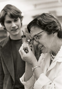
The windowless room, dank an dark, was not an obvious place for inspiration. I took notes, wondering if I would be able to glean anything meaningful from Professor Helen Stafford’s (1922-2011) meandering lecture. I was skeptical. After all, this was the same teacher who, annoyed with our choice of vegetarianism, had told us that “plants have feelings, too”.


But what I learned that day, 33 years ago, would trigger a grand curiosity about the natural world and draw me into the greatest scientific puzzle of my career.


Helen informed us that human language is not the only way that species communicate. Plants form intimate associations with fungi and bacteria, which allow them to thrive in stressful environments. Establishment and maintenance of the relationship depends on the passing and receiving of coded information between partners. She also told us that plants can only defend themselves against microbes that they can sense.

This interspecies communication is not restricted to plants and microbes. The human intestine is home to diverse bacteria, allowing us to harvest nutrients that would otherwise be inaccessible. The human immunodeficiency virus chooses for its target only those of us that carry a specific receptor, decorated in a particular way.

All these interactions dramatically affect human health and farm productivity.

I was hooked.

The Papers

This is the story behind the story of our two papers published last month in Plos One and Discovery Medicine.
We showed that single-celled bacteria communicate with each other using coded messages to coordinate attacks on their targets. Until now, the diversity of codes employed by these invading bacteria was thought to be extremely limited. We showed that one group of bacteria communicate by a novel, previously undescribed signal – and, as if to prove Helen Stafford correct- some plants have evolved a complementary cypher-breaking detection system that intercepts this bacterial code and then uses the information to trigger a robust immune response, preventing disease.

The Recent Background

The author Carol Shields has said that “The scientific life is the most complex of all to write about. In the case of scientists, impulse becomes compulsion”. And so it was with me. A year after college, I moved to Sweden to study with Professor Nils Fries who had identified a compound in pine trees that could coax some fungi to establish a lasting, mutually beneficial relationship. But pine trees and fungi and are difficult to study because they grow slowly. For this reason, when I returned to the US, I shifted my research to bacterial-plant interactions and their impact on plant health.

Rowland Biffen

In 1905, British geneticist and plant breeder Rowland Biffen demonstrated that it as possible to generate wheat varieties with resistance to a devastating diseases by moving genes around. He cross-pollinated a resistant wheat variety with a susceptible wheat variety and showed that the resulting seed carried the resistance of the parent.


Today, more than 100 years after Biffen’s discovery, plant breeders have introduced “resistance genes” into virtually every crop plant that we consume. But what are these genes? How do they function to detect the disease-causing microbe? For many years, the molecular basis of resistance remained elusive.

Brian Staskwicz

These were central questions addressed by the laboratory of Brian Staskwicz, where I carried out my PhD work. He had recently identified a bacterial protein that triggered an immune response in infected plants. He later identified one of the first plant resistance genes. After graduating, I moved to the laboratory of plant breeder Steven Tanksley at Cornell who was pioneering gene mapping in plants using molecular methods.

Focusing on Xa21


Figure 2.

My focus was a genetic locus, called Xa21, described in 1977 by Drs. S. Devadath, Gurdev Khush and collaborators. Rice plants carrying Xa21 have an unusual property: they are resistant to all known races of the bacterial pathogen Xanthomonas oryzae pv. oryzae (Xoo), which normally caused a devastating disease of rice in Asia and Africa. In 1992, I mapped this locus to a specific region in the rice genome. I hypothesized that it must consist of a cluster of tightly linked genes each recognizing a single race of the pathogen or a single gene that encoded a receptor recognizing a conserved microbial signature present in all races. When I moved to UC Davis I began a map-based cloning approach to isolate Xa21.


In 1995, two talented postdoctoral fellows in my lab, Guoliang Wang and Wenyuan Song, successfully isolated Xa21 and showed that it encoded a single gene: a receptor kinase. The structure immediately suggested that the XA21 protein could detect a microbial molecule present outside the plant cell and that this perception would activate an immune response inside the cell.

Toll and Toll-Like Receptors – Similar to XA21 – get the Nobel Prize in 2011

A few years after the discovery of the first plant resistance genes, the fly Toll and mouse Toll-like receptor (Tlr4) genes were isolated and shown to have striking structural similarities to XA21. Like XA21, TLR4 is membrane bound extracellular receptor that was predicted to bind a conserved microbial signature. TLR4 also carry the Toll /IL-1 Receptor (TIR) domain found in fly TOLL, the tobacco N resistance gene and the flax L6 resistance gene.

Thus, the discovery of a role for Toll and TLR4 in immunity provided a structural link between sensors utilized by plants and animals to detect infection. Professors Bruce Beutler and Jules Hoffman were awarded the 2011 Nobel Prize in Physiology or Medicine for their important work.


[Bruce and I share more than an interest in science; my father (Robert Rosenthal) and Bruce’s father (Ernst Beutler) were young cousins in Berlin in the 1920 and early 1930s. Their families fled the Nazi’s and reunited in the US after the war. I was honored to hear Bruce discuss XA21/Ax21 and our shared family history during his Nobel lecture last month (starts at 40:45)]


Characterizing Ax21


The next challenge was to isolate the conserved microbial signature produced by Xoo that was recognized by XA21.

Figure 4. RaxB

To isolate this molecule, which we named Ax21 (Activator of Xa21-mediated immunity), we screened for bacterial mutants altered in their ability to produce active Ax21. In this way, we isolated and characterized eight genes required for Ax21 activity (rax genes). raxA, raxB and raxC encode components of a predicted type I secretion system. Ax21 requires this RaxABC system for activity and secretion. The RaxB protein carries two highly conserved domains characteristic of proteins in Gram-positive bacteria that cleave N-terminal peptides prior to secretion of small proteins. Another set of rax genes encoded proteins important for sulfation.


These data suggested that Ax21 was a small sulfated protein with an N-terminal leader peptide, which was cleaved by the RaxB transporter prior to secretion outside the bacterial cell.

Much to our delight, that is exactly what we found.

Ax21 is a small protein with a tyrosine sulfation site and a predicted N-terminal leader sequence. Ax21 is not secreted in the absence of the RaxABC type 1 system. XA21/Ax21 binding triggers XA21-mediated innate immunity.

The Ax21 sequence is conserved in all Xanthomonas spp., in Xylella fastidiosa and the human pathogen Stenotrophomonas maltophilia. This conservation suggested that Ax21 serves a key biological function.

In our latest paper, we demonstrated, using two different methods, that the predicted Ax21 leader is cleaved as predicted. We further showed that only mature Ax21, missing the leader, is found outside the cell. When we add the purified protein back to a mutant lacking the wild-type gene, we can restore all Ax21 functions. These experiments demonstrate that the Ax21 protein acts outside the cell.  We then used a genetic approach to reveal that the likely bacterial receptor for Ax21, is a histidine kinase called RaxH. Finally, we show that the Ax21 mature protein has some pretty impressive capabilities: bacteria use it “talk” to each other.

Figure 1

Over the last 20 years, researchers have shown that bacteria employ specific signals to communicate. These signaling molecules, called “bacterial Esperanto” by Professor Bonnie Bassler, an early pioneer in studies of bacterial communication, accumulate in the external environment as the cells grow. When the concentration reaches a certain threshold level, the bacteria mobilize together to carry out concerted, group actions. This process is called quorum sensing.


Until now, it was thought that the two major groups of bacteria (Gram-positive and Gram-negative) use distinctly different types of communication codes. However, Ax21 doesn’t fall into either class. While the previously characterized signals in the bacterial coding repertoire were all relatively small molecules, Ax21 is a small protein, which makes it much larger.

Perception of Ax21 by the RaxH receptor triggers a massive change in the bacterial genetic program, altering the expression of nearly 500 genes, or approximately 10% of the bacteria’s genome. These changes allow the bacteria to assemble into elaborate protective bunkers, called biofilms, which render the bacteria resistant to dessication and antibiotic treatment. Thus, by virtue of communication and communal living, bacteria increase their chances of survival and proliferation. Ax21 perception also regulates the production of a virulent arsenal, including “effectors” that are shot directly into the host to disrupt its defenses and that initiate motility, allowing the bacteria to colonize new sites for infection.

Most rice plants are virtually defenseless against this Ax21-mediated bacterial attack – except for those plants that carry the XA21 immune receptor. This early detection gives the plant time to mobilize its defenses and mount an early and potent immune response.


S. maltophilia

The discovery that a small protein from a Gram-negative bacterium has a dual role in bacterial communication and in activation of the host innate immune response has not previously been demonstrated. We do not, however, believe this is an anomaly or that the biological importance of Ax21 is restricted to plant pathogens. We previously reported that Ax21 is also conserved in the nosocomial pathogen S. maltophilia and proposed a similar role for Ax21 in this species. Consistent with our hypothesis, a synthetic Ax21 protein has now been shown to regulate gene expression, motility, and biofilm formation in S. maltophilia, extending our findings to an animal pathogen.


Exploration of other bacterial genomes reveals the presence of an abundance of small secreted proteins similar to Ax21, suggesting the intriguing possibility that other species of bacteria also use small proteins to communicate and coordinate infection.

Control of Gram-negative bacterial infections in plants and animals remains a major challenge for the medical profession and for farmers, because conventional approaches are often not sufficient to eradicate these infections. One major reason for their persistence seems to be the capability of most bacteria to grow within biofilms that protect them from adverse environmental factors and antibiotics. The knowledge that bacteria use Ax21 to communicate can be used to develop reagents to immunize hosts against infection or antagonists to disrupt Ax21-mediated virulence activities and biofilm formation, a process thought to be involved in 65-80% of bacterial infections of plants and animals.

A Note on the Publication Process

Convincing the scientific community that a Gram-negative bacterium uses a small, N-terminal processed protein for bacterial communication has not been easy.

We initially submitted the manuscript to Science in 2009. One year, two manuscript versions, seven reviews, 16 figures and 11 tables later, we were still revising.

All but one of the six reviews were enthusiastic: they found the work “highly interesting”, the experimental work of “high quality”, carried out “to an exceptionally high standard” and the “evidence compelling”. The initial set of experiments the reviewers asked us to carry out were challenging but reasonable. We completed them over 7 months of intensive experimentation. Happily, the reviewer said we had “addressed all the fundamental issues” requested in the first round of reviews and that “the revisions were satisfactory”. Most of the additional suggestions were easily addressed. None of the reviewers challenged our central conclusion: that the Ax21 protein processed and secreted outside the cell where it serves as a quorum sensing factor. Indeed it is almost impossible to envision a different interpretation of these results.

However there was a problem.

One of the new reviewers pointed out that extracellular Ax21 must traverse the outer membrane to bind to the candidate RaxH receptor, which was predicted to localize to the inner membrane. The reviewer therefore asked that we isolate the genes encoding the proteins that make up the protein complex that imports Ax21. Such experiments, however, would require months, if not years, of additional work. Although we agreed that the requested set of experiments were interesting (and were already underway), successful completion would clearly not alter the fundamental conclusions of the paper. In addition, although the editor was kindly willing to consider our response to reviews, the editor cautioned us that the next version of the paper would be sent out to a THIRD set of reviewers.

(With my academic compulsion for accuracy, I initially wanted to include the second set of reviews, verbatim, as part of this post. Conversations with colleagues and the editor convinced me otherwise. They felt it would rupture an important agreement: that the conservations between editor, reviewers and author would remain confidential).

Because of the work described in the paper had already been talked about in public forums and included in grant applications, and because publication was important for moving forward with our grant applications, job applications and other papers, we felt we could not spend another year in the review process. The very essence of the scientific process is to challenge paradigms and share the experimental details with other scientists who can then reproduce or refute the findings. Publication is key for this process. We needed to publish.

After much discussion in the lab and several more discussions with the editor at Science, we decided to take advantage of the explosion of open access journals as an alternative route to publication.

I asked the editors at PLoS One if they would consider an expedited review. After reading the 4 science reviews, they generously agreed. They sent the paper out for an eighth review, which also came in positive. Finally, the paper was accepted. It was a long road but we are very glad the work is now available to other scientists to test and tweak. We hope it will be widely read.

As one distinguished colleague pointed out, ultimately it may not matter where the results are published; if it has legs, it will stand.


ResearchBlogging.org Han, S., Sriariyanun, M., Lee, S., Sharma, M., Bahar, O., Bower, Z., & Ronald, P. (2011). Small Protein-Mediated Quorum Sensing in a Gram-Negative Bacterium PLoS ONE, 6 (12) DOI: 10.1371/journal.pone.0029192

Press release from De Gruyter regarding acquisition of Versita re #OpenAccess

This press release may be of interest to those interested in Open Access publishing so I am posting it here:


De Gruyter acquires Versita and becomes third-biggest international Open Access publisher

Berlin, January 9, 2012
De Gruyter, the Berlin-based academic publishing company, is acquiring the publisher Versita. As a service provider to academic organizations and bodies, Versita publishes over 230 journals on Open Access basis, i.e. outside the traditional subscription model. With this acquisition De Gruyter is substantially increasing its presence in an important future market of academic publishing. The complete staff of Versita is being retained in this take-over.
“The purchase of Versita is a logical continuation of our publishing strategy of recent years, which involves giving both our authors and our customers an optimum range of publishing options”, says Dr. Sven Fund, the Managing Director of De Gruyter. “For us, the most important issue is that our publications have high-quality content, and not how they are financed.”

As of 2012 De Gruyter will be merging the newly acquired Open Access journals with its traditional subscription-based and freely sold content on one electronic platform, thus providing researchers with an outstanding service: users will have a powerful search interface for searching all academic articles from journals, books and databanks in which the articles have been published, independently of the various business models.

“For some time now, Open Access has been an important element of academic publishing. With the integration of all Versita content under a joint publishing administration on the new De Gruyter platform, we are now realizing the principle in our program,” continues Fund.
In the future, De Gruyter’s Open Access activities will be managed by Jacek Ciesielski as Vice President Open Access. Ciesielski founded Versita in 2001 and has been expanding the enterprise ever since. “Now that we have joined forces with De Gruyter, I look forward to further developing and internationalizing my activities together with De Gruyter”, comments Ciesielski.
Contact:
Ulrike Lippe
Manager Public Relations
De Gruyter: The independent academic publishing house De Gruyter can look back on a history spanning over 260 years.  The publishing group with headquarters in Berlin and Boston annually publishes over 800 new titles in the humanities, medicine, science and law and more than 230 journals and digital media. http://www.degruyter.com/
Versita publishes about 250 own and third-party scholarly journals across many disciplines. Its journal portfolio takes in 25 subscription-based journals, including a group of Central European Journals in 8 disciplines. Versita also publishes over 230 society journals, which are available through Versita Open (www.versitaopen.com), one of the largest Open Access platforms in the world. The company was established in 2001 by Jacek Ciesielski.http://www.versita.com/

Draft post cleanup #11: Tree Hugging

Yet another post in my “draft blog post cleanup” series.  Here is #11 from September.

Just a quick one.  In August a nice review paper came out on phylogenetic analysis software: Learning to Become a Tree Hugger | The Scientist.  By Amy Maxmen it is a “A guide to free software for constructing and assessing species relationships”.  Definitely worth checking out.

Among the key links & tools discussed:

Draft post cleanup #10: trip to LA artificially sweetened by Carolyn de la Peña

Yet another post in my “draft blog post cleanup” series.  Here is #10:

Went on a mini trip to UCLA for a mini meeting in November. It seemed appropriate that I brought with me to Los Angeles, land of empty pleasures – the new book from UC Davis Professor Carolyn de la Peña – “Empty Pleasures” on the history of artificial sweeteners. So I took a picture of the book overlooking part of LA from my hotel room:


The book is great read by the way …